|
|
Abstract |
|
|
|
|
High Resolution TEM Imaging with Hollow-cone illumination Maxim V. Sidorov, M. R. McCartney, and David J. Smith* Center for Solid State Science, Arizona State University, Tempe, AZ
85287-1704 It has was long been realized that imaging with hollow cone illumination (HCI) should, in theory, improve the directly interpretable resolution of TEM by as much as 100% (at the expense of contrast). The principle of HCI was first proposed by Scherzer 1 in 1949 and then reinvented by Hanssen and Trepte in 1971.2 As opposed to axial illumination, HCI effectively eliminates zeros and reversals on the transfer function providing direct interpretability of the resulting images.3,4 In addition to the substantial resolution enhancement, HCI should reduce significantly the phase-contrast noise inherent in axial HRTEM images.5 However, there are experimental obstacles for high resolution HCI which make its practical application very difficult to implement. To our knowledge, all observations using HCI so far have not shown all of the expected improvement predicted theoretically. This is believed to be due to the fact that accurate coma-free alignment is required to substantially improve the resolution.5 Another practical concern is the reduced contrast of the HCI images which can make it difficult to separate the useful signal from the background noise. Being inspired by a recent publication of Geipel and Mader,6 and by the latest advances in TEM instrumentation, we have implemented the HCI technique on our Philips CM200 FEG TEM. This microscope provides high enough coherence of the illumination to image spatial frequencies well beyond the point-to-point resolution limit (2.3 ). HCI was realized by manually setting a fixed tilt angle and continually changing the azimuth angle with a PC interfaced to the microscope using the remote control capabilities of the CM200. The resulting images were digitally recorded using a Gatan 794 CCD camera, allowing for subsequent image processing to improve/enhance the contrast. The improvements in image interpretability and resolution that might be available theoretically with our microscope are demonstrated in Fig. 1, which compares the appearance of <110> CdTe in standard HRTEM (a) and HCI (b) imaging modes. The individual atomic columns are clearly resolved with different contrast levels in the HCI mode.
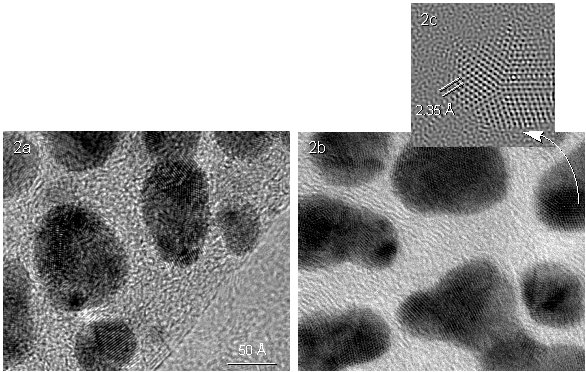
The practical application of the HCI technique to imaging of crystalline materials is shown in Fig. 2, which compares HRTEM images of a standard test sample (small Au particles on a thin C substrate) taken with axial (a) and hollow-cone (b, c) illumination. The HCI image displays significant image quality improvement not only due to the resolution enhancement but also by reducing the phase-contrast background noise.
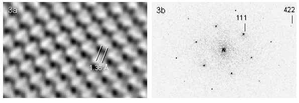
FIG. 2 HRTEM images of Au particles obtained with axial (a) and hollow-cone (b, c) illumination The significant resolution improvement obtainable with HCI is illustrated in Fig. 3a where Si dumbbells are directly resolved in the <110> projection. The Fourier transform of the HCI image of <110> Si is shown in Fig. 3b demonstrating information transfer up to, at least, the 1.095 [422] Si spot.
FIG. 3 Fragment of experimental hollow-cone image of <110>Si (a) where Si dumbbells are resolved, and Fourier transform (b) of the image It is worrying that the image characteristics are varying so rapidly across the small field of view in Fig. 3a: possible reasons, which are under investigation, include changes in local crystal tilt, and the effect of three-fold astigmatism and non-isoplanicity of the illumination across the field of view. Moreover, complete image compensation of the image shift during image recording has not yet been achieved. Nevertheless, it is encouraging that these preliminary results demonstrate, at least partially, the expected improvements in resolution and image interpretability.7 References
|
|
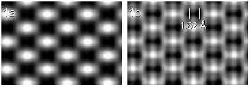 FIG. 1 Simulated HRTEM images of <110> CdTe for axial (a) and
hollow-cone (b) illumination
FIG. 1 Simulated HRTEM images of <110> CdTe for axial (a) and
hollow-cone (b) illumination