Presentation

Up

High Resolution TEM Imaging with Hollow-cone illumination

presented at MSA'97 Meeting, Cleveland, OH
August 13, 1997

[Abstract is here]

Imaging with tilted illumination:
how does it work?

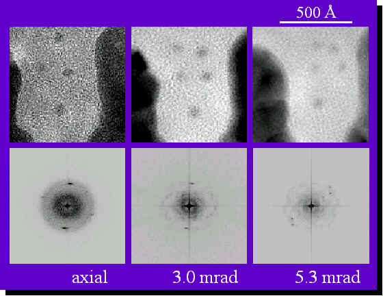
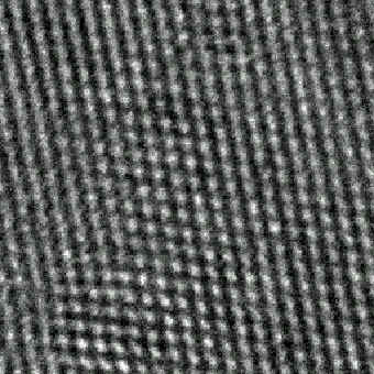
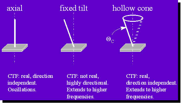
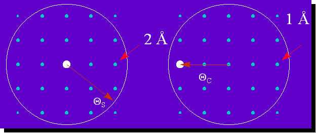
For axial illumination (above, left) the incident beam is in the center (in reciprocal space) and directly interpretable information transferred through the microscope is limited either by the objective aperture or by point-to-pint resolution of the microscope (if no objective aperture is used). In the case shown above, information up to 2 gets transferred.
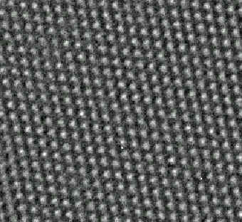
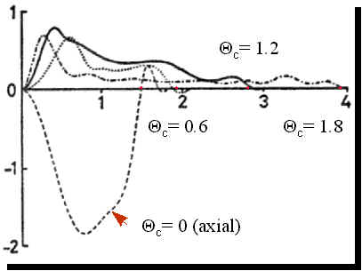
Now, if one tilts the beam as shown above (right), 1 information gets transferred. In the case of fixed tilt, this resolution enhancement will be highly directional and, in fact, in the perpendicular direction one would lose resolution.
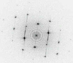
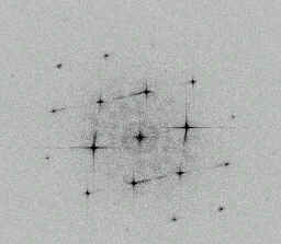
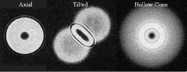
Hollow cone illumination takes care of this directionality as can be seen in the picture below where 2-dimensional contrast transfer functions corresponding to axial, fixed tilt, and hollow cone are shown.


Experimental Images
Gold particles on amorphous carbon

axial and hollow-cone (for two different tilt angles) images
Note: reduced contrast from amorphous carbon

axial (left)and hollow-cone (right) images
Note: nicely resolved Au lattice and reduced contrast from amorphous carbon

Si/SiO2 interface

axial (bottom) and hollow-cone (top) images
Note: nicely resolved Si lattice and reduced contrast from amorphous SiO2

hollow-cone image of Si (right)
Note: Si dumbbells are clearly resolved!
Left: Fourier transform of the image
Note: presence of the 422 peak of Si indicates information transfer up to (down to?) 1.095

CdTe

axial image of CdTe and its Fourier transform

hollow-cone image of CdTe and its Fourier transform

High-pass filtered hollow-cone image of CdTe
Note CdTe dumbbells in the inset


Last updated: April 28, 2017

 
home.gif (1097 bytes)Go to Max Sidorov Web
1996-2017 Maxim V. Sidorov, All Rights Reserved